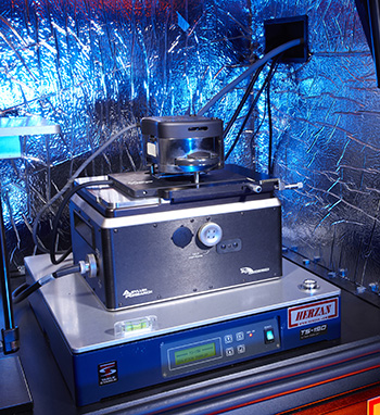
The Atomic Force Microscope (AFM) belongs to the family of scanning probe microscopes (SPM). It consists of a cantilever, typically silicon, with a sharp tip at its end which is scanned very precisely over the specimen surface with piezo-electric motors. When the tip is brought into proximity to a sample surface, forces between the tip and the sample lead to a deflection of the cantilever following Hooke's law, which is precisely measured with a laser.
The versatility of the technique regarding materials analysis relies on the fact that a large variety of forces can be used to induce the deflection of the cantilever. Depending on the situation it is possible to use van der Waals, electrostatic, magnetic, capillary, solvation, and chemical bonding forces, among many others. Correspondingly the technique can be applied to a wide range of materials, from very soft to very hard, from conductive to non-conductive, etc. Furthermore, it is possible to use specialized tips that allow measuring simultaneously additional quantities such as local temperature, thermal conductivity, heat capacity, and so on.
Spatial resolution is strongly influenced by the shape of the tip. For flat samples, the lateral resolution depends on its radius of curvature. With the adequate tip, it is in principle possible to achieve atomic resolution. Nevertheless, with standard tips, nanometer scale observations are more realistic. Vertical sample variations can be measured with sub-nanometer resolution. For large aspect ratios, it has to be considered that the actual surface topography is modulated by the tip shape.

Asylum MFP-3D Standard System with Low Force Indenter
Operation Modes
Contact and tapping (AC) mode; lateral force mode (LFM); phase imaging; electric force microscopy (EFM); nanolithography; force curve mode; ramp mode; force mapping mode.
Features
90 micron travel in (x, y) and 15 micron in z; X-Y closed loop (non-linearity <0.5 %) with sensitivity < 150 pm noise; Z-closed loop (<0.2 %) with sensitivity < 35 pm noise; ARgyle-3D imaging in real time; AFM control program integrated with IGOR pro.
Asylum MFP-3D BioApplication |