Focused ion beam (FIB) microscopy operates similarly to a scanning electron microscope (SEM) in that a fine beam of in this case Gallium ions is scanned over the surface of the material, generating secondary signals in form of electrons or ions which can then be used to visualize the samples' morphology. However, due to the high ion mass, the beam additionally sputters material off the surface providing the opportunity to expose sub-surface structures. The ion beam can be focused down to a few nanometers allowing extremely site-specific ablation of material. By simultaneously flowing specific gases over the sample, it is possible to either enhance the etch rate or conversely deposit materials such as platinum, tungsten, or carbon.
Modern dual beam systems combine SEM and FIB in one instrument. The ion beam can efficiently remove volumes in the order of several ten-micron cubed. By doing this in a controlled fashion the material can be exposed slice by slice and correspondingly imaged using various secondary signals with the SEM. A three-dimensional volume rendering is then achieved by stacking the 2D data (serial sectioning).
FIB has become a quintessential tool in modern transmission electron microscopy due to its unique ability to extract TEM samples from very specific regions of the material. The technique can be applied to a wide variety of materials. Modern ion columns can adequately focus ion beams with energies down to 1keV opening the opportunity to significantly reduce the surface damage introduced by gallium ion implantation. As a result, high-quality samples thinner than 50 nm can be prepared which are accessible to atomic-resolution microscopy.
Another important application is the preparation of samples for atom probe tomography (APT, see chemical analysis tools section), which makes use of the capability to extract material, transport it to a specific holder and apply circular milling patterns to prepare a 100 nm thin needle with a tip radius of curvature of a few ten nanometers.
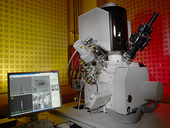
FEI Helios Dual-beam 600 FIB/SEM
A high-resolution dual-beam focused ion beam and scanning electron microscope. The instrument is equipped with an Elstar field emission SEM for nanometer resolution imaging and a Tomahawk FIB column operating from 30 kV down to 500 V. Ion currents range from 7 pA, providing a minimal beam size of 7 nm, to 22 nA which can be used for substantial milling tasks. The very stable piezo-driven stage can operate on an area of 150 x 150 mm. Attached is currently a gas source for platinum deposition as well as an Omniprobe manipulator that is typically used for the transfer of TEM and APT samples.
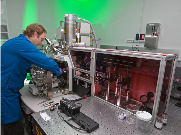
Tri-Beam Laser/FIB/SEM
Dual-beam focused ion beam and scanning electron microscope equipped with a femtosecond laser and associated scanning and focusing optics. The laser can be used for rapid serial sectioning of large volumes, combined with multimodal imaging and spectroscopy using SEM, electron back-scattered detection, and electron dispersive spectroscopy.